Disturbing images explain why we don’t MRI pregnant women
Topics: Social Media, Science, Health, Parenting
 Britt Jones
Topics: Social Media, Science, Health, Parenting
In what is probably the creepiest image on the internet comes a lesson as to why we don’t make pregnant women go through a magnetic resonance imaging (MRI) machine.
You might have seen a few pictures on social media that set your teeth on edge, but this will give it a run for its money.
There has not been anything more awful to see since the MRI pug fiasco online, until now.
2022 was a terrifying time to be on X, as when @dhomochameleon shared three images of what looked to be a spooky pickle, people took notice.
Advert
Some wondered what they were looking at, others wanted to scrub it from their memories forever, but mostly, people were shocked at what it was.
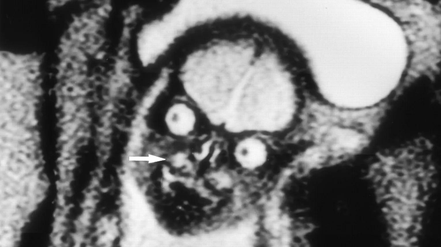
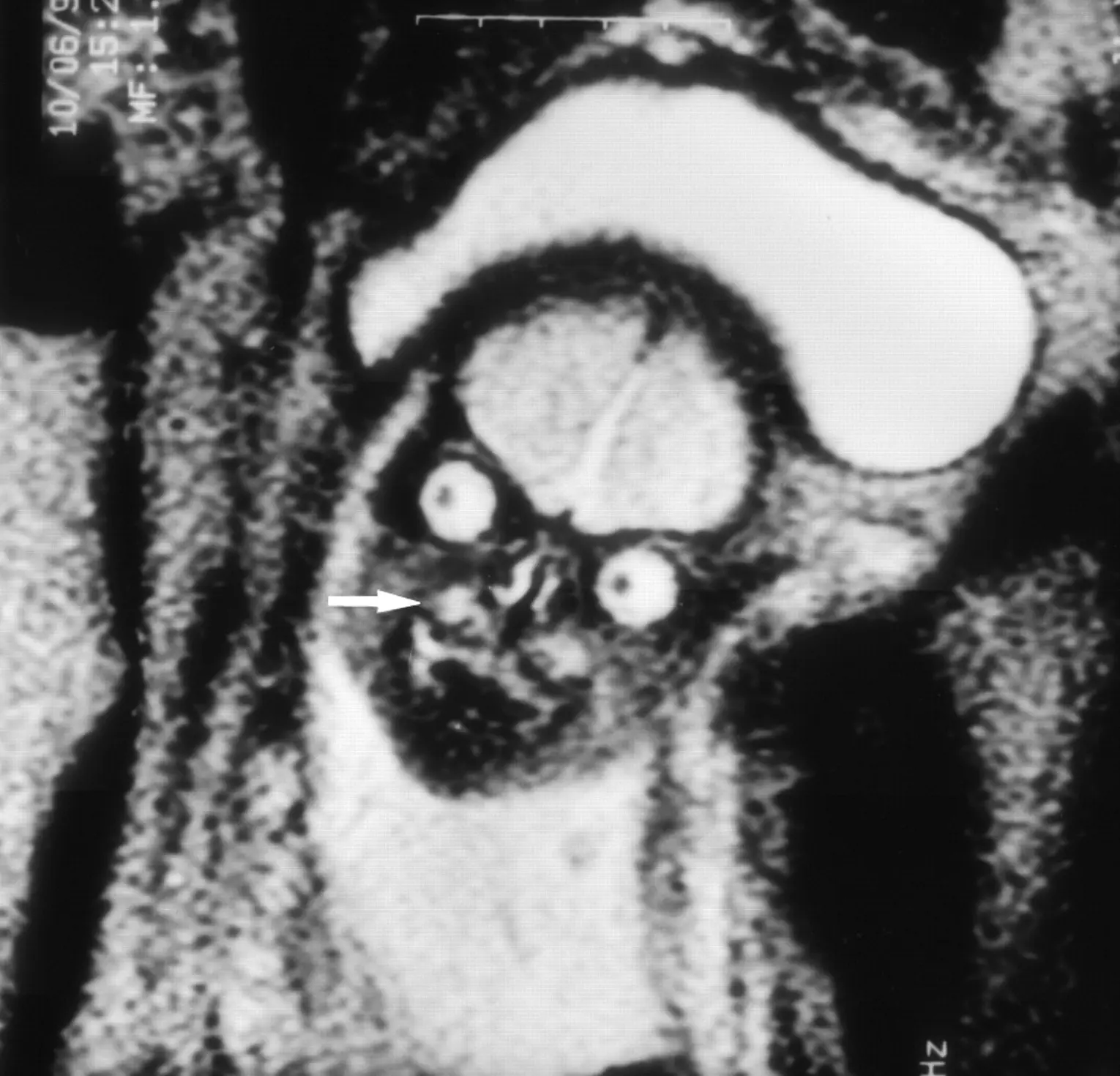
A feotus inside its mothers' stomach.
Snopes interviewed Jason Moody, a graduate research assistant at the University of Wisconsin, who shockingly confirmed the pictures were real, and that researchers at Harvard Medical School had similar images on file when studying the brains of babies.
As it turns out, two of the images shared to X had also been shared by two Harvard researchers of feotuses at 19 and 24 weeks of gestation.
You might be asking why the feotuses look like goopy jack-o-lanterns or that creepy thing from Mars Attacks, but there’s actually a very simple reason.
"MRI makes it very easy to differentiate between different types of soft tissue found in the body and we are mostly soft tissue. Remember, we are mostly water," Moody said. "One of the primary features of these images are the substantial signal differences between the eyes, brain, nose, and the rest of the face."
The ‘giant magnet’ sees things no other machine can, which takes you to a strange layer of the human body.
To do it, Moody explained how they image mothers, noting: "We place a coil of wire around patients as the protons in their body rotate and we use that coil to measure the energy released by the rotating protons as a current. That current is then converted into an image and different tissue types will look differently from one another based on how differently their protons behave (how quickly they lose energy)."
It gives a super detailed view of a baby’s brain before they are born, in cases where they are worried about its development or need to double check things.
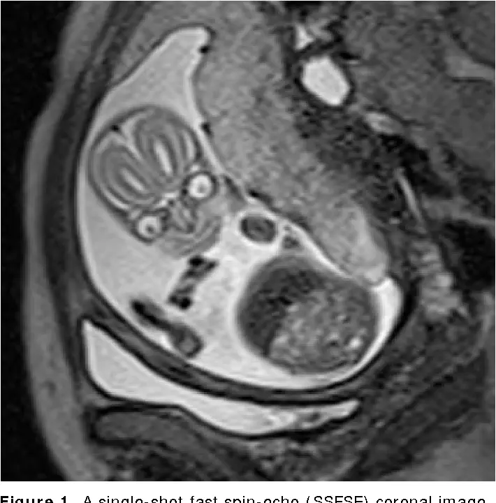
According to an article published by RSNA in its RadioGraphics issue, advances in magnetic resonance (MR) imaging have almost eliminated the need for fetal premedication, because they can now see clearly the baby in utero via their technology.
Similarly, a 2013 neuroimaging study published in Semantics Scholar also confirmed that the MRI can find things that an ultrasound is unable to detect due to its level of penetrative viewing.
For those online, it is a horror to behold...but also funny.
“Scary yes but hilarious af,” said one.
The real reason they discourage MRIs during pregnancy is because then people would realise they’re incubating nightmare demons and would be rightfully terrified pic.twitter.com/55zEeOofsP
— Katie (@ZiziFothSi) May 19, 2021
“Eeeeyyyyuuucckkkk,” said another.
While those pictures alone should be enough to sway anyone against prescribing an MRI session to a pregnant mother, it's not the reason why they are so uncommon.
Per Radiology Info, t's because they are typically reserved for the second and third trimester of the pregnancy, and generally, they just aren't usually needed if an ultrasound is sufficient in picking up easy to see abnormalities.
It's when things are hard to see, or need to be seen via the magnet imaging that they'd issue an appointment.